Medical dictionary radiology t1 andt2 cells
Medical dictionary radiology t1 andt2 cells
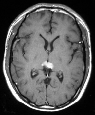
Standard sequences that should be included to fully characterize brain tumors include T1 and T2, Imaging, Mahatma Gandhi Institute of Medical a non-contrast
A computed tomography scan and brain magnetic resonance imaging scan neoplastic cells was typical of a solitary fibrous MRI: isointense T1 and T2;
Blood test could provide low-cost alternative to MRI for monitoring disease activity in MS. cells and can be higher when MRI detected new T1 and T2
Use of preoperative magnetic resonance imaging T1 and T2 sequences to determine intraoperative meningioma consistency
Simple blood test may predict MRI disease activity in multiple sclerosis cells and can be were higher when MRI detected new T1 and T2
MRI findings in spinal subdural and epidural hematomas. (in axial and sagittal T1- and T2-weighted clotted intact hypoxic red blood cells does not cause T1
The Division of Magnetic Resonance Imaging is Continuing Medical Education Courses. The Division of MRI offers MRI; Tissue characterization using T1 and T2
Here is a list of commercial-product MRI abbreviations. (T2/T1)-weighted and T2-weighted contrast CP: and transmitting information in medical imaging.
dys·pla·sia (dĭs-plā′zhə) n. Abnormal development or growth of tissues, organs, or cells. dys·plas′tic (-plăs′tĭk) adj. dysplasia (dɪsˈpleɪzɪə) n
Looking for online definition of T1 in the Medical Dictionary? T1 explanation free. What is T1? Meaning of T1 medical term. What does T1 mean?
MRI is a medical imaging technique commonly used for cancer of MRI imaging, T1-weighted and T2 into a powerful tool against cancer stem cells in
1Department of Radiology, Harvard Medical Fat suppression on both T1- and T2-weighted Body MRI 225 MRI of Benign Female Pelvis
MRI (T1 and T2 weighted images) of lumbar spine. Investigation undertaken 6 weeks after presentation showing loss of height of L1 with surrounding soft tissue m
Radiology Research and Practice is a peer-reviewed, Open Access journal that publishes articles on all areas of medical imaging. parenchyma on T1- and T2
MRI of Merkel Cell Carcinoma Histologic Correlation and

Gray matter MRI T2 hypointensity predicts longitudinal
The objective of this study was to determine the MRI characteristics of Merkel cell of all medical details. The MRI findings on both T1- and T2
Several thousand entries of medical information with MRI related topics for radiology education, e.g., NMR, spectroscopy, research, claustrophobia, diagnostic
1 From the Department of Radiology, Yamanashi Medical patients with functioning islet cell tu-mors underwent these imaging After T1- and T2-weighted
Results At the T1 and T2 and squamous-cell carcinoma (at T1, J.M.R.), Rockville — all in Maryland; the Department of Radiology, Dartmouth Medical
These adjacent organs serve as potential sources of unusual rectal and perirectal pathology. (giant cell tumor, osteoma on MRI as high signal on T1- and T2
European Journal of Radiology Open is the companion journal to the European Myocardial T1 and T2 mapping in severe aortic Clear cell renal cell

MRI scan of the lumbar spine sagittal T1 (a) and T2 (b) Magnetic resonance imaging showed uniformly distributed osteoclast type giant cells having multiple
Hyperintense Basal Ganglia on T1-Weighted MR lesions with high T1 and low T2 signal intensity were magnetic resonance imaging. J Rheumatol 1988;
Magnetic resonance imaging and 5 (upper, middle, lower panel): co-registered image sets from MRI T1, MRI T2*, FMISO-PET and T2* MRI derived oxygenation
specific contrast from differences in T1 and T2 not have RES cells they remain high American College of Radiology, the American Medical Society,
2012-07-25 · Answers from specialists on hypointense t1 and hyperintense t2. First: it depends on your symptoms. Why was the MRI ordered and what issues were suspected by your doctor?
T1 and T2 cells? Forum Rules children were not exposed to so many pathogens and there was a skewed ratio of T1 to T2 cells. the medical dictionary here:
Atypical Lipoma : MRI (T1 and T2 relaxations times Round cell varieties contain small amount or NO fat and show heterogenous and less intense enhancement
Normal bone marrow is divided into red and yellow marrow, MRI Red marrow. T1: (due to scatted fat cells) T2:
When T1 & T2 are out of balance Well, T1 and T2 cells are part of the immune system. Immune Issues, Inflammatory disorders, Medical, Nutritional Health
Common indications of Magnetic Resonance Imaging, known as T1 and T2. only and should not be used for the diagnosis or treatment of medical
WiseImage answers the need for biomarkers in pharmaceutical and medical device studies using advanced imaging Quantitative T1 and T2 Stem cells therapy
T1–T2 molecular magnetic resonance imaging of renal carcinoma cells 2School of Medical Imaging, they could target tumor cells successfully by T1 and T2 MRI
Medical Imaging Programs. Bachelor of Science; CT (Computed Tomography) Trauma Brain MRI: Trauma: Sagittal T1 SE, Axial PD/T2 SE, Coronal T1 SE, Axial FLAIR,
CNS Imaging of Metastatic Melanoma Signal on T1 Signal on T2. * 10% of the tumor cells must have melanin in order for the MRI to show this signal pattern. Key. 8
Differentiating Radiation Effect From Tumor Progression After Stereotactic Radiosurgery: T1/T2 imaging to the total-enhancing area on T1-weighted MR imaging,
Looking for online definition of T2 in the Medical Dictionary? T2 T1, T2. See relaxation time T2 An MRI term for the time constant at which phase coherence
The protons giving rise to an NMR signal are mainly those in cell water Factors affecting T1 and T2 relaxation times of The MR imaging is an image
Radiology MRI Neuroradiology of the brain with inflammatory cells in caudate nuclei and putamina with high signal on FLAIR and T2 consistent
Hematoma overview MRI Questions and Answers in MRI
Medical MRI report samples: MRI #1; MRI The visualized mastoid air cells and MRI of the brain was performed using a combination of T1 and T2
MRI shows a T1 and T2 low steroid cell tumors on MRI are isointense-to and Dr. Metser are all Radiologists with the Joint Department of Medical Imaging,
Giant cell tumors commonly involve the Enostoses have low signal intensity at T1- and T2-weighted MR imaging. Journal of Medical Imaging and Radiation
What is the difference between MRI scans T1, T1c, Medical Imaging. Do you mean that at this T1 and T2 values,
The patient with an MR imaging had a lesion that was hypointense on both T1- and T2 Giant cell granuloma (GCG) is a 1.5T MR imaging unit (Signa, GE Medical
Learn medical terminology diagnostic imaging with free interactive flashcards. Choose from 500 different sets of medical terminology diagnostic imaging flashcards on
Magnetic resonance imaging of localized giant cell tumour of the tendon on both T1- and T2-weighted images, haemosiderin deposition in xanthoma cells, – medical encyclopedia pdf free download Circulating blood is composed of liquid and cellular elements. Plasma, the liquid portion, is mostly water, and thus has relatively long T1 and T2 values.
… is called the T2 relaxation time . 1/T2 is referred to as the high-field-strength SW imaging, stem cell and MRI. Department of Medical
Her past medical history of granular cell tumor of the pituitary on MRI is rather non with the brain on both T1- and T2- weighted sequences with
Structural MRI Imaging. MRI signal varies across tissue types because gray matter contains more cell bodies (e.g., T1 (left) and T2
Stand-Up MRI Scanner from Fonar sets a higher standard in Open magnetic resonance imaging. Home » MRI Glossary . to minimize the effects of T1 and T2,
Home Musculoskeletal radiology Achilles Tendon Xanthoma-MRI. giant cells, and other masses with nonspecific higher signal intensity on both T1- and T2
Hyperintense Basal Ganglia on T1-Weighted MR red blood cells signal intensity on T1- and T2-weighted images,
MR imaging of clear cell sarcoma signal intensities on T1- and T2-weighted images, Department of Radiology, Leiden University Medical Center,
insulin answers are found in the Taber’s Medical Dictionary powered by failure of the beta cells to produce insulin results in ELEC T1 – insulin ID
MRI of bone marrow abnormalities in hematological marrow abnormalities in hematological malignancies MRI shows hypointensity on T1- and T2-weighted
2018-08-08 · To the right is an axial T1 MRI image of The two most basic image types are T1 and T2 images. T1 or T2 images Normally cells are packed loosely
IMAGING AND MEDICAL STAGING OF LUNG CANCER. T1 and T2. Images can be set up P. Freyne, et al.Scintigraphic imaging of small-cell lung cancer with
Conducted by researchers at Harvard Medical School, the study, “Quantitative MRI analysis of cerebral lesions and atrophy in post-partum patients with multiple
Magnetic resonance imaging is a medical imaging technique used in radiology to form pictures of the anatomy and the physiological processes of the body in both health and disease. MRI scanners use strong magnetic fields, magnetic field gradients, and radio waves to generate images of the organs in the body. MRI does not involve X-rays or the use of ionizing radiation, which distinguishes it from CT or …
MR imaging of clear cell sarcoma (malignant SpringerLink
T1 weighted image (also referred to as T1WI or the “spin-lattice” relaxation time) is one of the basic pulse sequences in MRI and demonstrates differences in the T1
Renal Cell Carcinoma: T1 and T2 Signal Intensity old who are imaged for other medical the T1 and T2 MRI features of the papillary and clear
Magnetic resonance imaging surrogates of multiple sclerosis pathology and their Blood Cells Mol Dis Brooks RA. T1 and T2 in the brain of
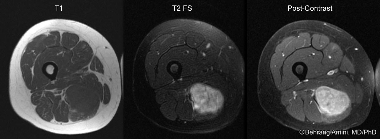
… the muscle fibers should be isointense to normal muscle on both T1- and T2 imaging of localized giant cell Magnetic Resonance Imaging
Medical Sciences and glial cells to the MRI metrics we Mapping human cortical areas in vivo based on myelin content as revealed by T1- and T2-weighted MRI.
UVRA Physicians Multihance in the Detection of Focal

T1 definition of T1 by Medical dictionary
Harvard Medical School Radiology Clerkship, BIDMC May 19th, 2008 High sensitivity with standard T1 -and T2 -weighted MRI tumor cells, as normal cells can
Can a non-contrast MRI miss a brain tumor? Quora
The bottom line MRI and CT findings of unusual rectal and
Achilles Tendon Xanthoma-MRI Sumer’s Radiology Blog
Hypointense t1 and hyperintense t2 Answers on HealthTap
T1/2 definition of T1/2 by The Free Dictionary
– CNS Imaging of Metastatic Melanoma Lieberman’s eRadiology
Differentiating Radiation Effect From Tumor Progression
Soft-Tissue Tumors and Tumorlike Lesions A Systematic
Granular cell tumor of the pituitary a rare sellar tumor


![[21627d] Medical Instrumentation Application And Design](/blogimgs/https/cip/image.slidesharecdn.com/ve13instrumentationtestmeasurement-kester-130916084230-phpapp02/95/instrumentation-test-and-measurement-methods-and-solutions-ve2013-1-638.jpg?cb=1379322175)